Central Nervous System Diagram - Nervous System Mepedia
Central Nervous System Diagram - Nervous System Mepedia. Figure 12.8a functional and structural areas of the cerebral cortex. Create healthcare diagrams like this example called central nervous system in minutes with smartdraw. The cns receives input from a variety of different sources, and implements an appropriate response to the stimuli, in a cohesive manner. The nervous system monitors and controls almost every organ system through a series of positive and negative feedback loops.the central nervous system (cns) includes the brain and spinal cord. It controls the voluntary movements, balance of the body.
ads/bitcoin1.txt
Figure 12.8a functional and structural areas of the cerebral cortex. The central nervous system consists of the brain and spinal cord. Gustatory cortex (in insula) primary motor cortex Central nervous system function coordination and movement. Some reflex movements can occur via spinal cord pathways without the participation of brain structures.
Central Nervous System An Overview Neupsy Key from neupsykey.com The pns connects the cns to the rest of the body. Central nervous system (cns) | central nervous system parts | structure and function of central nervous system | central nervous system diagram biologist april 16, 2021. The cns consists of two organs which are continuous with each other; Finally, some nerves are mixed nerves that contain both afferent and efferent axons. Central nervous system function coordination and movement. Start studying central nervous system. Smartdraw includes 1000s of professional healthcare and anatomy chart templates that you can modify and make your own. The central nervous system (cns) is the largest part of the nervous system.it is made up of the brain and the spinal cord.
Related posts of central nervous system diagram anatomy of human liver.
The central nervous system (cns) and the peripheral nervous system (pns).the central system is the primary command center for the body, and is comprised of. The brain plays a central role in the control of most bodily functions, including awareness, movements, sensations, thoughts, speech, and memory. In this section, we focus on the peripheral nervous system; A thin thread called filum terminale extends from the tip of the conus medullaris all the way to the 1st coccygeal vertebra (co1) and anchors the spinal cord in place. We explore the types of cells involved, the regions of.
How The Peripheral Nervous System Works from www.verywellmind.com The central nervous system consists of the brain and spinal cord. But some scientists have classified them into two divisions in which the ans is included under peripheral nervous system category. Other neurons, known as efferent nerves, carry signals only from the central nervous system to effectors such as muscles and glands. Figure 12.8a functional and structural areas of the cerebral cortex. Anatomy of human liver 10 photos of the anatomy of human liver anatomy human body liver, anatomy human digestive system, anatomy human heart, anatomy human kidney, anatomy human lungs, anatomy human pancreas, anatomy human stomach, inner body, anatomy human body liver, anatomy human digestive system, anatomy human. Peripheral nervous system collections of neuron cell bodies associated with nerves in the peripheral nervous system are known as __________. Some reflex movements can occur via spinal cord pathways without the participation of brain structures. The diagram at the right.
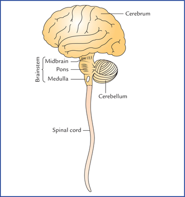
Central and peripheral nervous system diagram 2 / 10 ( 1 vote ) in this image, you will find central and peripheral nervous system, brain, frontal lobe, parietal lobe, occipital lobe, temporal lobe, midbrain, pons, medulla, cerebellum, spinal chord, autonomic, somatic, parasympathetic, sympathetic in it.
ads/bitcoin2.txt
The brain interpretes the messages and despatch them to the effectors through efferent (motor) neurons. The nervous system, essentially the body's electrical wiring, is a complex collection of nerves and specialized cells known as neurons that transmit signals between different parts of the body. Put simply, the cns is the supreme command center of the body. We explore the types of cells involved, the regions of. It gathers information from all over the body and coordinates activity. Other neurons, known as efferent nerves, carry signals only from the central nervous system to effectors such as muscles and glands. Figure 12.8a functional and structural areas of the cerebral cortex. Central nervous system (cns) | central nervous system parts | structure and function of central nervous system | central nervous system diagram biologist april 16, 2021. Trascina e rilascia le puntine sull'immagine, nella posizione corretta. The central nervous system (cns) consists of the brain and the spinal cord, while the peripheral nervous system (pns) consists of sensory neurons, ganglia (clusters of neurons) and nerves. Multiple sclerosis (ms) is a potentially disabling disease of the brain and spinal cord (central nervous system). Nerves that carry information from sensory receptors to the central nervous system only are called afferent nerves. Some reflex movements can occur via spinal cord pathways without the participation of brain structures.
The peripheral nervous system (pns) concerns all the nervous system outside the central nervous system and contains motor and sensory nerves which transmit information to and from the body and brain. Multiple sclerosis (ms) is a potentially disabling disease of the brain and spinal cord (central nervous system). The cns receives input from a variety of different sources, and implements an appropriate response to the stimuli, in a cohesive manner. In ms, the immune system attacks the protective sheath (myelin) that covers nerve fibers and causes communication problems between your brain and the rest of your body. The central nervous system is made up of the brain and spinal cord.
209 Central Nervous System Illustrations Clip Art Istock from media.istockphoto.com It controls the voluntary movements, balance of the body. The cns consists of two organs which are continuous with each other; Anatomy of human liver 10 photos of the anatomy of human liver anatomy human body liver, anatomy human digestive system, anatomy human heart, anatomy human kidney, anatomy human lungs, anatomy human pancreas, anatomy human stomach, inner body, anatomy human body liver, anatomy human digestive system, anatomy human. The diagram at the right. In ms, the immune system attacks the protective sheath (myelin) that covers nerve fibers and causes communication problems between your brain and the rest of your body. The central nervous system (cns) and the peripheral nervous system (pns), shown in.the cns is comprised of the brain and spinal cord; But some scientists have classified them into two divisions in which the ans is included under peripheral nervous system category. The central nervous system (cns) and the peripheral nervous system (pns).the central system is the primary command center for the body, and is comprised of.
The nervous system has two major parts:
ads/bitcoin2.txt
Learn vocabulary, terms, and more with flashcards, games, and other study tools. The central nervous system (cns) is the largest part of the nervous system.it is made up of the brain and the spinal cord. Impulses are transmitted to the central nervous system through afferent (sensory) fibres of the peripheral nerves. It controls the voluntary movements, balance of the body. Related posts of central nervous system diagram anatomy of human liver. Later, we look at the brain and spinal cord. Gustatory cortex (in insula) primary motor cortex The brain interpretes the messages and despatch them to the effectors through efferent (motor) neurons. Smartdraw includes 1000s of professional healthcare and anatomy chart templates that you can modify and make your own. The cns receives input from a variety of different sources, and implements an appropriate response to the stimuli, in a cohesive manner. The brain is the central information processing organ of our body and act as the 'command and control system'. A thin thread called filum terminale extends from the tip of the conus medullaris all the way to the 1st coccygeal vertebra (co1) and anchors the spinal cord in place. It gathers information from all over the body and coordinates activity.
ads/bitcoin3.txt
ads/bitcoin4.txt
ads/bitcoin5.txt
0 Response to "Central Nervous System Diagram - Nervous System Mepedia"
/what-is-the-peripheral-nervous-system-2795465-FINAL-b69e1bb803654212a83d9e68eb4847d0.png)


0 Response to "Central Nervous System Diagram - Nervous System Mepedia"
Post a Comment